 | –≠–ª–µ–∫—Ç—Ä–æ–Ω–Ω—ã–π –∫–æ–º–ø–æ–Ω–µ–Ω—Ç: C7876 | –°–∫–∞—á–∞—Ç—å:  PDF PDF  ZIP ZIP |

NEW
The C7876 series X-ray image intensifier (X-ray I. I.) camera
units are ideal for non-destructive inspection of light-element
materials and radiation imaging at low-energy X-ray levels. The
C7876 remarkably improves X-ray transmittance at low-energy
X-ray levels because a beryllium (Be) window is used instead of
the aluminum (Al) window currently used in most X-ray image
intensifiers.
The result is a clear, distinct image taken in real time even at
low-energy X-ray levels down to several keV. For example, the
internal structure of thin resin or aluminum objects, which have
been difficult to visualize, now can be clearly observed with high
contrast.
q
X-ray transmittance of window materials
4" X-ray Image Intensifier with Beryllium Window
Efficiently Coupled to CCD Camera
X-RAY I.I. CAMERA UNIT
C7876, C7876-10
Captures low-energy X-ray images in real time!
v
Flower
(X-ray tube voltage: 10kV)
TII B0097EA
0.5 mm
AI
0.5 mm
Be
1
10
100
90
80
70
60
50
40
30
20
10
0
X-RAY ENERGY (keV)
X-RAY TRANSMITTANCE (%)
100
The C7876 series is an X-ray image intensifier camera unit
using a 4" X-ray image intensifier with a beryllium window,
efficiently coupled to a built-in high-sensitivity CCD camera.
OVERVIEW
qIdeal for X-ray imaging at low-energy levels
(for thin resin, aluminum objects, etc.)
qHigh resolution, high contrast
qLow distortion
FEATURES

BERYLLIUM WINDOW EFFECTS
The C7876 series uses an X-ray image intensifier having a beryllium (Be) window instead of the aluminum (Al) window
commonly used in most X-ray image intensifiers. This has brought a significant improvement in X-ray transmittance at low-
energy X-ray levels, making it possible to capture a clear, high contrast X-ray image of objects which up until now have
been difficult to capture with good contrast.
v
Glass fiber reinforced plastics
(X-ray tube voltage: 16kV, tube current: 100
µ
A)
v
Glass fiber reinforced plastics
(X-ray tube voltage: 16kV, tube current: 100
µ
A)
C7876 CONTROLLER
OFFSET
GAIN
ENHANCE
POWER
ON
OFF
C7876 DC POWER SUPPLY
POWER
ON
OFF
TII C0060EA
Unit Configuration: X-Ray I. I. Head CCD Controller DC Power Supply for X-Ray I. I.
CABLE LENGTH: 5m
CABLE LENGTH: 5m
DC Power Supply
Controller
X-ray Image Intensifier Head
HIGH-VOLTAGE
POWER SUPPLY
X-RAY IMAGE
INTENSIFIER
X-RAY
SOURCE
OBJECT
Optional 10-meter cables are also available.
100 - 240 V
25 VA
100 - 240 V
10 VA
CCD
CONFIGURATION
Aluminum Window 0.5mmt
With an aluminum window, no contrast can
be obtained in the X-ray image of an object
like plastics, but with a beryllium window ....
Beryllium Window 0.5mmt
With a beryllium window, the fiber structure
can be clearly viewed with high contrast.
IMAGING EXAMPLES
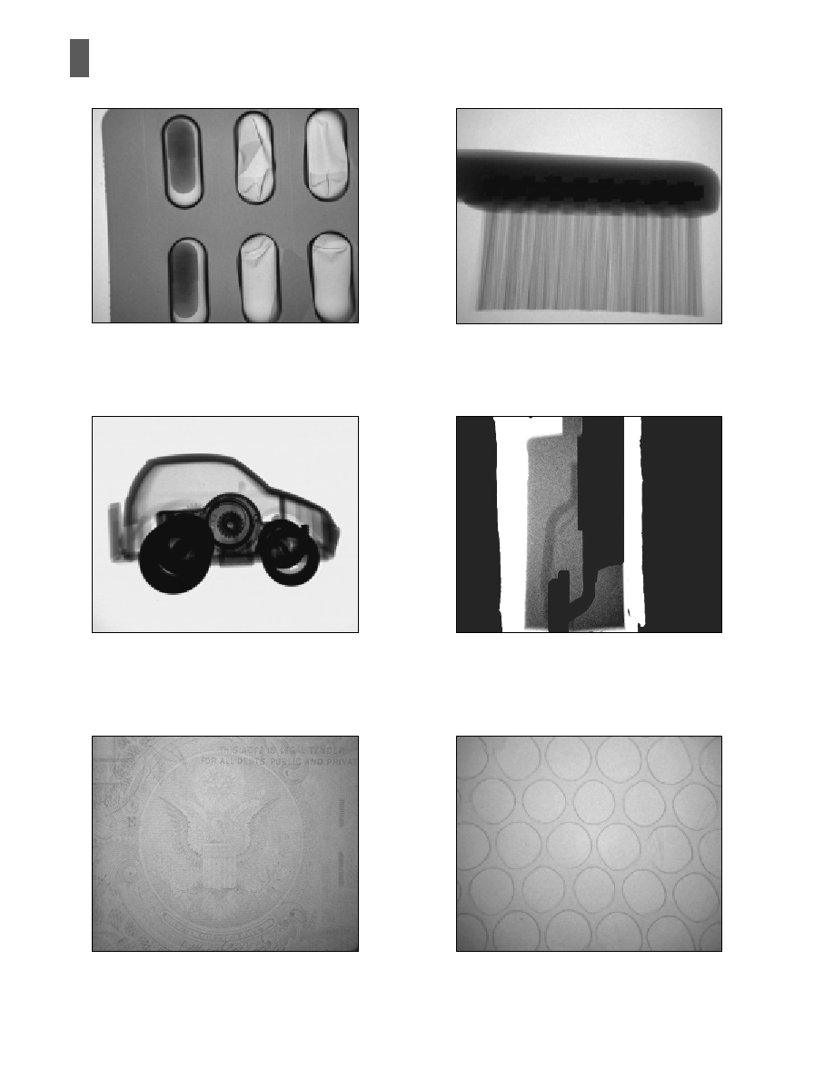
v
Drug capsules
(X-ray tube voltage: 16kV)
Drug capsules can be checked for proper
filling or cracks in the aluminum film.
v
Toy
car
(X-ray tube voltage: 20kV)
The outline of a plastic car body and the
internal spring winding can be distinctly
observed simultaneously.
v
Dollar bill
(X-ray tube voltage: 9kV)
Ink printed on the front and back surfaces can
be viewed clearly.
v
Toothbrush
(X-ray tube voltage: 18kV)
The internal body structure and individual
nylon bristles can be observed clearly.
v
Aluminum bonding wires
(X-ray tube voltage: 30kV)
Aluminum bonding wires difficult to visualize
with a conventional X-ray I. I. (aluminum
window) can be viewed with high contrast.
v
Air cushioning material
(X-ray tube voltage: 10kV)
Slight differences in thin film thickness can be
viewed with high contrast.

Parameter
Input Window Material
Input Phosphor
Output Phosphor
Imaging Area (on input surface)
Resolution
A)
(on input surface)
Scanning Method
CCD
Signal
Processing
Functions
Input Voltage
Power Consumption
Weight
Beryllium (0.5mm
t
)
CsI
P-20 or equivalent
72 (H)
54 (V) average
4.6 average
NTSC/BW
CCIR
2/3" FIT 410,000 pixels
2/3" FIT 480,000 pixels
4 : 3
61
Sync composite video signal 1.0Vp-p/75
Gain control range: 1 to 30 times
Offset control range: 0 to -100%
Horizontal and vertical parabolic correction:
±
30%
Horizontal and vertical slant correction:
±
30%
0 to 9
1 / 0.45
100 to 240 (50/60Hz)
35
Approx. 8
Approx. 3
--
--
--
mm
lp/mm
--
--
--
dB
--
--
--
--
--
dB
--
Vac
VA
kg
kg
Unit
C7876-10
C7876
CCD Chip
Aspect Ratio
S/N ratio
Output Signal
Enhancement Function
Shading Correction
AP Correction
Gamma Correction
Head
Controller & DC Power Supply
A) Measured with an optimum tube voltage, using an X-ray resolution chart directly fitted to the X-ray image intensifier.
NTSC B/W : National Television System Committee
CCIR : Comite Consultatif Internationale des Ratio Communications
SPECIFICATIONS
BERYLLIUM WINDOW SAFETY PRECAUTIONS
Beryllium (Be) is used for the window of this product. Beryllium is solid under normal conditions and not
harmful as long as it stays unchanged in quality. However, if beryllium is mechanically or chemically pro-
cessed and inhaled as fumes or in powder form, it may be extremely harmful. To prevent beryllium from
entering your body through a cut, breathing, eating or drinking, NEVER TOUCH THE BERYLLIUM WINDOW
WITH BARE HANDS. Should you touch the beryllium widow or debris when broken, immediately wash your
hands or other parts of the body that came in contact with the beryllium.
Beryllium is designated as a chemical substance that must be handled in accordance with authorized
disposal methods. If this product is broken or has reached the end of its service life, please contact our sales
office to ship it back to us and we will take the proper means to dispose of it.
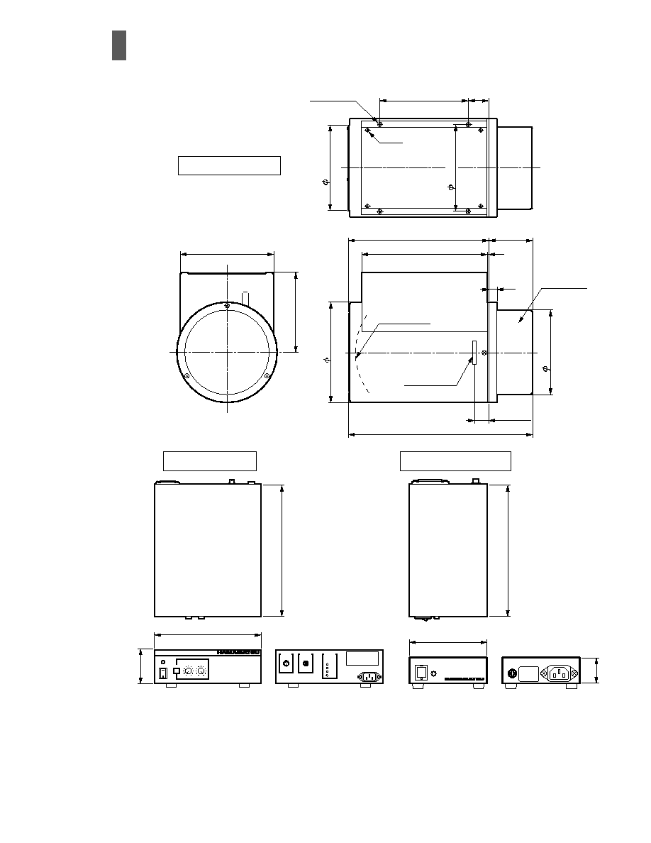
DIMENSIONS
(Unit: mm)
TII A0044EA
INPUT
PHOSPHOR
SCREEN
CAMERA
HEAD
HV POWER SUPPLY
OUTPUT
PHOSPHOR
SCREEN
2
14
226
±
2
71
±
1
205
±
2
164
±
2
126
±
2
297
±
3
25
±
0.3
154
±
1
130
±
2
130
±
1
4-M6 D15
120
±
0.5
42
4-M3
136
±
0.5
C7876 CONTROLLER
OFFSET
GAIN
ENHANCE
POWER
ON
OFF
CAMERA
VIDEO OUT
SHADING
AC IN
VP
VS
HP
HS
C7876 DC POWER SUPPLY
POWER
ON
OFF
AC IN
DC
OUT
110
220
280
180
70
40
Front View
Front View
Rear View
Rear View
X-ray I. I. Head
Controller
DC Power Supply
Subject to local technical requirements and regulations, availability of products in this promotional material may vary. Please consult with our sales office.
Information furnished by HAMAMATSU is believed to be reliable. However, no responsibility is assumed for possible inaccuracies and omissions.
Specifications are subject to change without notice. No patent rights are granted to any of the circuits described herein. © 1999 Hamamatsu Photonics K.K.
HAMAMATSU PHOTONICS K.K., Electron Tube Center
314-5, Shimokanzo, Toyooka-village, Iwata-gun, Shizuoka-ken, 438-0193, Japan, Telephone: (81)539/62-5248, Fax: (81)539/62-2205
U.S.A.: Hamamatsu Corporation: 360 Foothill Road, P. O. Box 6910, Bridgewater. N.J. 08807-0910, U.S.A., Telephone: (1)908-231-0960, Fax: (1)908-231-1218
Germany: Hamamatsu Photonics Deutschland GmbH: Arzbergerstr. 10, D-82211 Herrsching am Ammersee, Germany, Telephone: (49)8152-375-0, Fax: (49)8152-2658
France: Hamamatsu Photonics France S.A.R.L.: 8, Rue du Saule Trapu, Parc du Moulin de Massy, 91882 Massy Cedex, France, Telephone: (33)1 69 53 71 00, Fax: (33)1 69 53 71 10
United Kingdom: Hamamatsu Photonics UK Limited: Lough Point, 2 Gladbeck Way, Windmill Hill, Enfield, Middlesex EN2 7JA, United Kingdom, Telephone: 44(20)8-367-3560, Fax: 44(20)8-367-6384
North Europe: Hamamatsu Photonics Norden AB: Smidesv‰gen 12, SE-171-41 SOLNA, Sweden, Telephone: (46)8-509-031-00, Fax: (46)8-509-031-01
Italy: Hamamatsu Photonics Italia: S.R.L.: Strada della Moia, 1/E, 20020 Arese, (Milano), Italy, Telephone: (39)02-935 81 733, Fax: (39)02-935 81 741
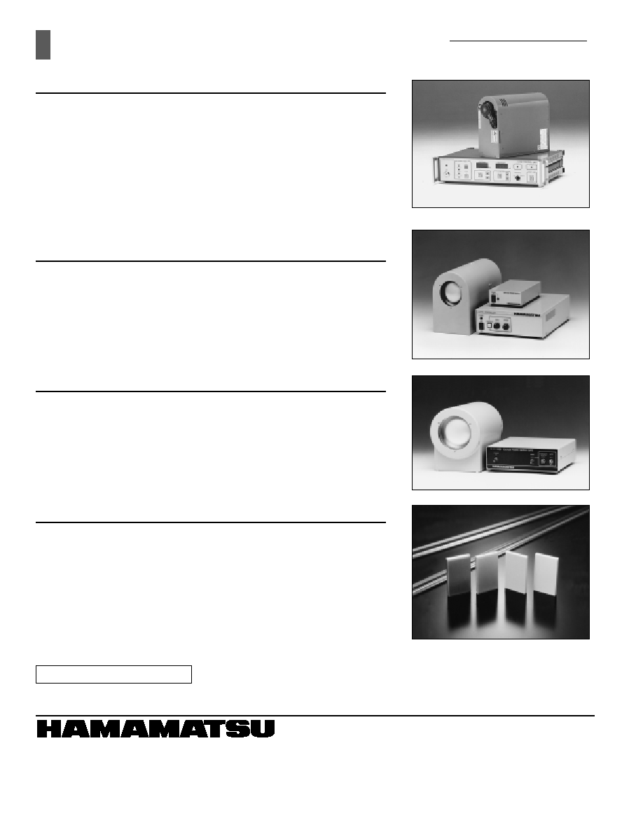
RELATED PRODUCTS
130kV Microfocus X-Ray Source
The Hamamatsu microfocus X-ray source uses an X-ray tube with an extremely
small focal spot of 10 microns in diameter. This gives a sharp, clear image even at
magnified image. When used with the C7876 series X-ray I. I. camera unit, high-
quality X-ray images can be taken in fine detail even under high magnification.
Besides the 130kV model, Hamamatsu offers various models of microfocus X-ray
sources, including 80kV, 100kV and 150kV models.
X-ray Image Intensifier Camera Unit (3"/1.8" Dual Mode)
C7716, C7716-10
The C7716 series X-ray image intensifier (I. I.) camera unit now offers greatly
improved X-ray detection efficiency even at low energy levels. This improvement
stems primarily from a built-in X-ray image intensifier having an extremely thin
aluminum input window. Its thickness is only 0.3mm or less, virtually at the limit of
present technology providing excellent X-ray transmittance and low scattering.
The results are sharp, clear, high-quality images taken at low energy X-ray levels
down to several keV which penetrate plastic (PET) materials.
X-ray Image Intensifier Gate Unit C7077
The C7077 X-ray image intensifier gate unit consists of a gateable 4-inch X-ray
image intensifier (X-ray I. I.) with a relay lens and a gate power supply. Since the X-
ray I. I. itself has a gating function, still X-ray images of a high-speed moving object
can be taken by inputting an external trigger signal while using a normal X-ray
source. This means there is no need for a pulsed X-ray source. Gating can be
performed by a TTL level trigger pulse, allowing easy gate control.
FOS (Fiber optic plate coated with X-ray scintillator)
The FOS is an optical device for X-ray imaging, fabricated by coating an X-ray
scintillator material over a fiber optic plate consisting of tens of million glass fibers
of several micrometers each in diameter. The FOS provides higher sensitivity and
resolution than currently used sensitized paper films and also allows real-time
digital radiography when directly coupled to a CCD. The fiber optic plate used in the
FOS has excellent X-ray absorption characteristics, so that X-rays which penetrate
the X-ray scintillator and directly enter the CCD are minimized to less than 1%. This
protects the CCD from "deterioration" and "noise increase" caused by X-ray
irradiation, assuring a long service life and maintaining high image quality.
X-RAY I. I. CAMERA UNIT
C7876, C7876-10
TII 1047E01
OCT. 1999 WT
Printed in Japan (500)
CE marking status underway